A Clearer Window Into Disease: Meet the Opera Phenix Microscope
At the root of every major disease of our time is the simple question: what is going wrong with...At the root of every major disease of our time is the simple question: what is going wrong with our cells? Once scientists know exactly how cells are getting sick, they can figure out how to prevent or stop the dysfunction and bring patients back to health.
Thanks to a very special microscope at the NYSCF Research Institute, this process is faster and more accurate than ever. The instrument, called the Opera Phenix, can show scientists exactly how diseased cells are behaving, what is wrong with them, and how we might be able to fix them – it can literally see things that are invisible to most labs – features that might hold therapeutic opportunities that others are missing. It can also capture cell images on a large scale, showing the diversity of large groups of cells that makes it possible to conduct detailed studies.
The Opera was gifted to NYSCF by longtime supporter and Leadership Council member Dorothy Lichtenstein, who hopes that it will enable exciting new discoveries to further personalized medicine.
For NYSCF researchers, the impact of this transformational equipment is immeasurable.
“The Phenix allows for truly automated imaging, taking human bias out of image acquisition. What separates it from other microscopes is the excellent autofocus capabilities, which match those of human users. Having the bottleneck of focusing out of the equation, we are left with amazing image quality that lets us study disease in unprecedented detail,” remarked Tom Rusielewicz, PhD, a Principal Scientist who oversees the Opera’s use.
“The Opera Phenix also gives us a higher-resolution view of structures within a cell that might be malfunctioning in the disease, which we can then target with new therapies,” he explained.
Dive into the Opera’s extraordinary images with us and learn about how they are accelerating disease research!
Seeing Stem Cells like Never Before
The image on the right shows induced pluripotent stem cells – stem cells made on the NYSCF Global Stem Cell Array® from a sample of skin or blood. The power of stem cells is that they can make unlimited copies of themselves, and scientists can tell here that these cells are beginning to multiply.
These stem cells can go on to become any type of cell in the body, all while carrying the unique genetic blueprint of the individual from whom they are derived. NYSCF’s world-leading technology allows researchers to create high-quality stem cells from hundreds of individuals at a time, changing the way diseases are studied.
A Close-Up on Cancer
Using the Opera Phenix, NYSCF scientists are tracking the growth of tumor organoids — living avatars of patient tumors created with the power of stem cells. No two tumors are the same, and this image shows organoids colored to categorize different characteristics.
[metaslider id=16590]
NYSCF scientists use these patient-specific organoids to improve the way women’s reproductive cancers are studied and to develop personalized treatments as part of our Women’s Reproductive Cancers Initiative.
“The Opera Phenix has allowed us to scale up our characterization and analysis of these unique models, illuminating the heterogeneity of the disease. Looking at different organoids across patients, as seen in the second photo, shows how the disease can manifest in variable ways, and grouping them helps scientists understand which types of patients are developing which disease features,” explained Laura Andres-Martin, PhD, a Research Investigator in Oncology who leads the Initiative.
Parsing Out the Drivers of Parkinson’s Disease
Parkinson’s disease (PD) is a tricky disease to study and treat because researchers do not yet fully understand what causes dopaminergic neurons – the cells affected by PD – to start deteriorating.
The left side of the image below shows dopaminergic neurons, the cells that are lost in Parkinson’s disease, derived from a healthy volunteer, and the right side shows microglia derived from a PD patient.
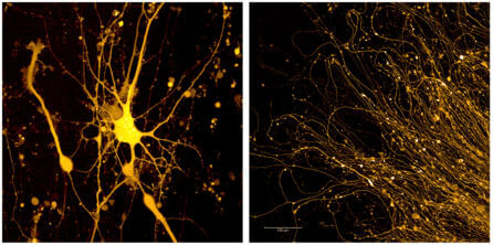
“Unlike the neurons derived from a healthy donor (which grow in a straight line), these patient cells grow in circles. This may give us clues as to why these cells are dying in Parkinson’s brains,” noted Angelique di Domenico, PhD, a postdoctoral fellow in Parkinson’s disease and neurodegeneration.
Brain cells other than dopaminergic neurons are likely involved in PD. The left side of this image shows microglia, the immune cells of the brain, derived from the stem cells of a healthy donor. Comparing these microglia to those derived from Parkinson’s patients, pictured on the right side of the image, gives us insight into what might drive the disease.
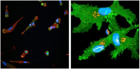
“Microglia move very fast, and the Opera Phenix can image them quickly without inducing damage as other microscopes would,” said Dr. Di Domenico.
In these images, scientists stained the microglia’s Golgi apparatus, a part of the cell which may be implicated in the disease, in yellow.
Investigating the Intricacies of Multiple Sclerosis

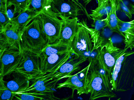
Multiple sclerosis (MS) is a complex disease involving multiple cell types in the brain. In MS, the immune system attacks myelin, the protective coating surrounding nerve fibers that helps them send signals. Myelin is made by cells called oligodendrocytes, and studying these cells may help researchers understand the root of the disease and develop new therapies.
The oligodendrocytes to the left were made from the stem cells of a healthy donor, and comparing them to oligodendrocytes derived from multiple sclerosis patients helps researchers understand how the disease develops.
“All of this complex and challenging image analysis is easily performed by the Opera Phenix’s software, which greatly speeds up our research,” said Maria Sapar, PhD, an Associate Staff Scientist who imaged the cells.
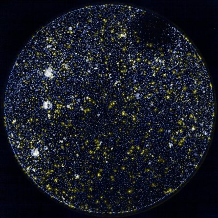
The image to the right shows oligodendrocytes derived from the stem cells of an MS patient. The white spots are cells that are showing a high signal (increased activity).
“Looking at patterns across groups of diseased cells can point to drivers of
disease,” noted Dr. Sapar. “The Opera Phenix can be set up to automatically find and image the cells we are interested in, speeding up data acquisition and analysis to accelerate scientific discovery”
A New View of the Cells that Make Vision Possible
The image below shows retinal pigment epithelial (RPE) cells, the eye cells that are depleted in age-related macular degeneration (AMD), leading to vision loss.
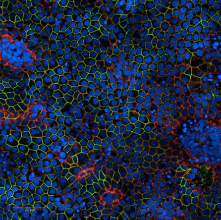
“These cells make up the equivalent of the blood-brain barrier for the eye. Using the Opera Phenix, we can see how these cells strongly tile together to create this barrier in the eye and how this structure becomes disrupted in macular degeneration,” noted Cecile Terrenoire, PhD, Senior Director of Process Development.
NYSCF recently completed construction of a facility for creating clinical-grade cells, including RPEs to be used in a cell therapy for AMD. For this treatment, scientists will turn stem cells from different AMD patients into RPE cells, and each patient will then receive a transplant of ‘their own’ RPE cells to restore their vision.
Imaging RPEs can also offer insight into why these cells begin to deteriorate in AMD in the first place. The image above shows RPEs stained to highlight different features that may be relevant to disease.
“Analyzing how RPEs from healthy donors differ from AMD patients allows researchers to understand what triggers the disease, how to reverse or prevent it, and how to develop healthy RPE cells for cell replacement therapies,” said Dr. Terrenoire.
Artificial Intelligence and the Opera: Seeing the Unseeable
By integrating artificial intelligence with NYSCF’s stem cell technology, our researchers can identify new features of disease and discover drugs that reverse these features. Recently, NYSCF scientists published a pre-print detailing how their unbiased, artificial-intelligence-driven platform – developed in collaboration with Google Research – is working to identify cellular characteristics of PD, opening the door for discovering effective drugs. Studies like this require large numbers of images to enable artificial intelligence, and the Opera greatly accelerates this work.
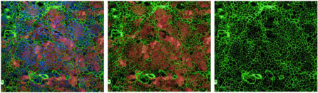
The images in the below slideshow were made by applying different fluorescent stains to the same group of cells to highlight different structures. NYSCF scientists are using artificial intelligence to examine these structures in incredible detail thanks to the Opera Phenix’s high resolution, detecting disease characteristics that would be imperceptible to the human eye.
[metaslider id=16613]