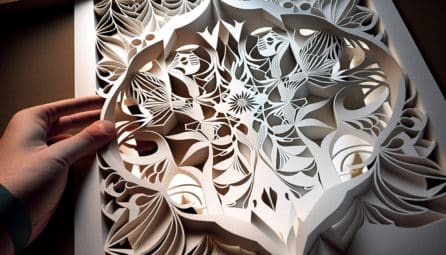
How An Extra-Intricate Type of Origami Inspired a New Window into the Brain
The Context: Brain organoids – 3D avatars of human brain tissue made from stem cells – are a revolutionary...The Context: Brain organoids – 3D avatars of human brain tissue made from stem cells – are a revolutionary tool for studying disease. However, it has been difficult for scientists to record electrical activity (a key component of brain function) from organoids because they are small and fragile.
The Study: Inspired by the Japanese art of kirigami, scientists led by NYSCF – Robertson Stem Cell Investigator Alumnus Sergiu Pasca, MD, of Stanford University have devised a flexible, hammock-like fabric of electrodes that can record electrical activity in brain organoids without damaging them. The study appears in Nature Biotechnology.
The Importance: This new tool will help scientists more accurately model the brain and record its electrical activity, giving important insights into development and disease.
This research had an unlikely birthplace (a cafe) and an unlikely muse (the ultra-intricate art of kirigami). It began when Dr. Pasca ran into fellow scientist, chemist Bianxiao Cui, PhD, at a Stanford coffee shop called Peet’s.
They began discussing their research, and Dr. Pasca expressed his latest challenges with organoids: specifically, to record their electrical activity without damaging them.
“As we began building neural tissues that were ever-more complex, the main limitation was not how we make them, but actually how we’re going to precisely probe and manipulate them,” said Dr. Pasca in an article from Stanford.
The problem is that most methods for monitoring electrical activity are built for brains in living organisms or on fixed surfaces, not for free-floating, miniature blobs of brain cells. Sometimes researchers will cut organoids in half and put them on an electricity-recording surface, but this doesn’t work so well either.
“In just a few days, they become like a pancake,” Dr. Cui. “They’re no longer free-floating, and lose some of their unique properties.”
Paper Folding Provides a Path Forward
Drs. Pasca and Cui have been aiming to develop a better method for years. It wasn’t until they took inspiration from the complex art of kirigami that they found their solution.
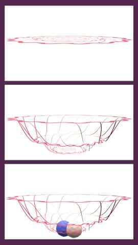
The scientists realized what they needed was more of a net than a solid surface – something that could contort itself to adjust to an organoid’s environment rather than the other way around. They engineered a woven fabric of electrodes, called ‘kirigami electrodes’, that does just this, acting as a kind of hammock that allows organoids to continue their natural development and can record electrical activity without inducing damage, and continuously.
“We’ve never been able to continuously monitor one neural organoid to see when electrical activity emerges,” remarked Dr. Pasca. “And that’s actually a fundamental question, even for understanding human brain development.”
What’s Next?
Next, Dr. Pasca hopes to expand the kirigami electrodes to see if they can be applied to assembloids, larger aggregates of tissues that mimic interactions between brain regions. He also notes that he’s excited to see how other scientists in the field may employ the tool in their own research.
“I think the power of this technology will be in how people use it.”
Journal Article:
Kirigami electronics for long-term electrophysiological recording of human neural organoids and assembloids
Xiao Yang, Csaba Forró, Thomas L. Li, Yuki Miura, Tomasz J. Zaluska, Ching-Ting Tsai, Sabina Kanton, James P. McQueen, Xiaoyu Chen, Valentina Mollo, Francesca Santoro, Sergiu P. Pașca. Nature Biotechnology (2024). DOI: 10.1038/s41587-023-02081-3